
Virtual Reality / Plant Science & Biotechnology
Internal Structure of Dicot Monocot Stem VR Simulation Training
Overview
Internal Structure of Dicot Monocot Stem VR Simulation Training by ASFAN gives learners focused, repeatable practice through immersive scenarios that support skill development, assessment, and safer preparation.
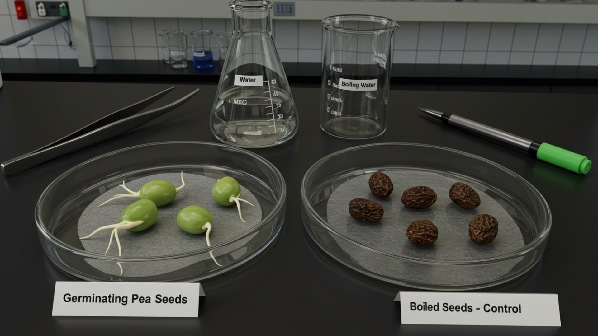
Learning Objectives
- Recognize and differentiate key anatomical features in stems and roots of dicots vs. monocots.
- Identify tissue layers: epidermis, cortex, vascular bundles (xylem & phloem), pith, endodermis, pericycle (for roots), etc.
- Observe spatial arrangement and pattern differences between dicot and monocot vascular bundles.
- Understand functional significance of each tissue type (support, transport, storage).
- Explore both longitudinal and cross-section views dynamically.
Key Benefits of the VR Experience
- Safe, no-prep alternative to real dissections-no microscopes, stains, or samples required.
- Interactive 3D learning improves spatial understanding beyond flat diagrams.
- Clear visual differentiation between dicot and monocot anatomy.
- Repeatable and accessible exploration anytime, anywhere.
- Great for remote learning or limited lab resources.
Virtual Lab & Sample Selection
A clean, well-equipped virtual lab with options to choose plant type (dicot or monocot) and to examine stem or root cross-sections.
Virtual Dissection & Sectioning
Use virtual tools to cut stems or roots, generate realistic cross-sections, and rotate, zoom, and pan to inspect layers from any angle.
Tissue Layer Visualization & Labeling
Clear rendering of epidermis, cortex, vascular bundles, pith, xylem, phloem, endodermis, pericycle (roots), with color/texture coding and interactive labels explaining each function.
Comparative Mode: Dicot vs. Monocot
Side-by-side comparison highlighting bundle arrangements (ring vs. scattered), presence/absence of pith, cortex thickness, root structure differences, and why these matter.
Exploration & Learning Tools
Zoom to cellular level, toggle labels or outlines, switch to microscope view, and optionally quiz yourself to identify tissues by name.
Explore More Projects
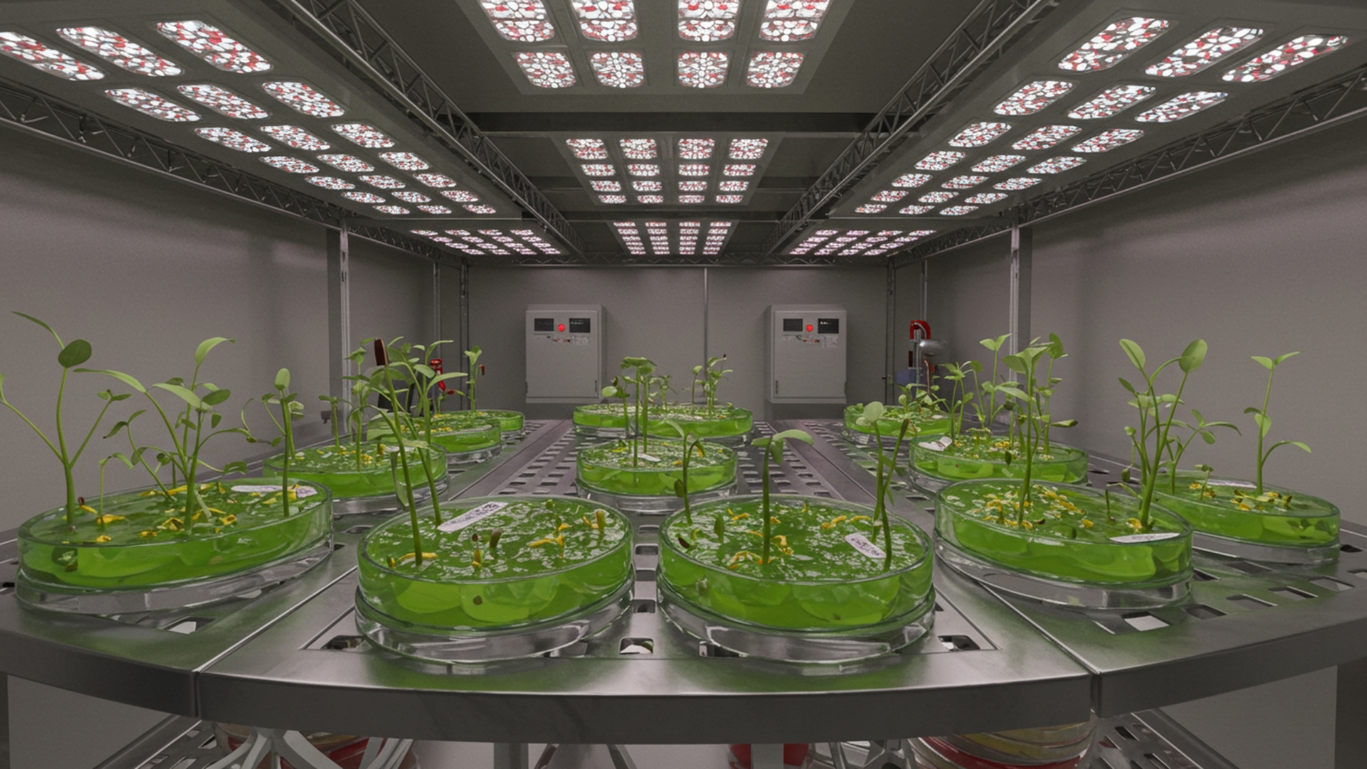
VR Micropropagation: Shoot Tip Culture
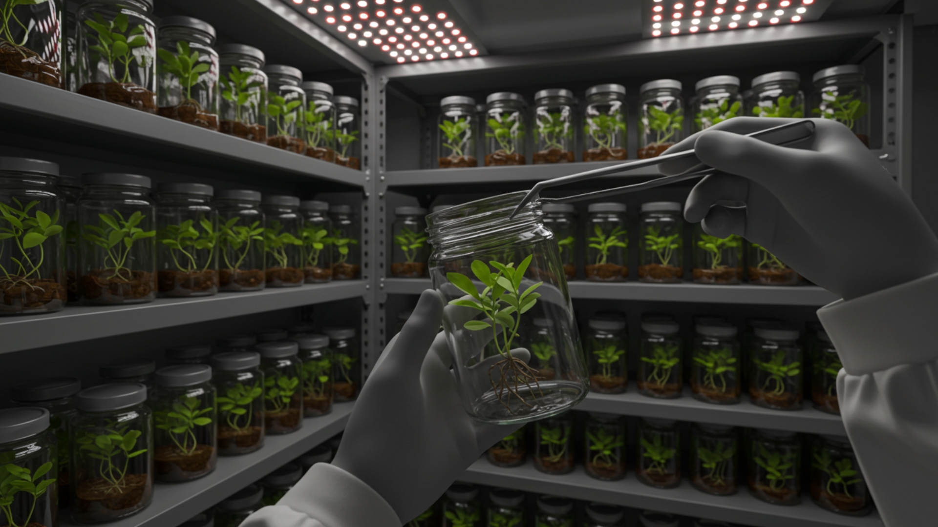
VR Micropropagation: Axillary Bud Culture
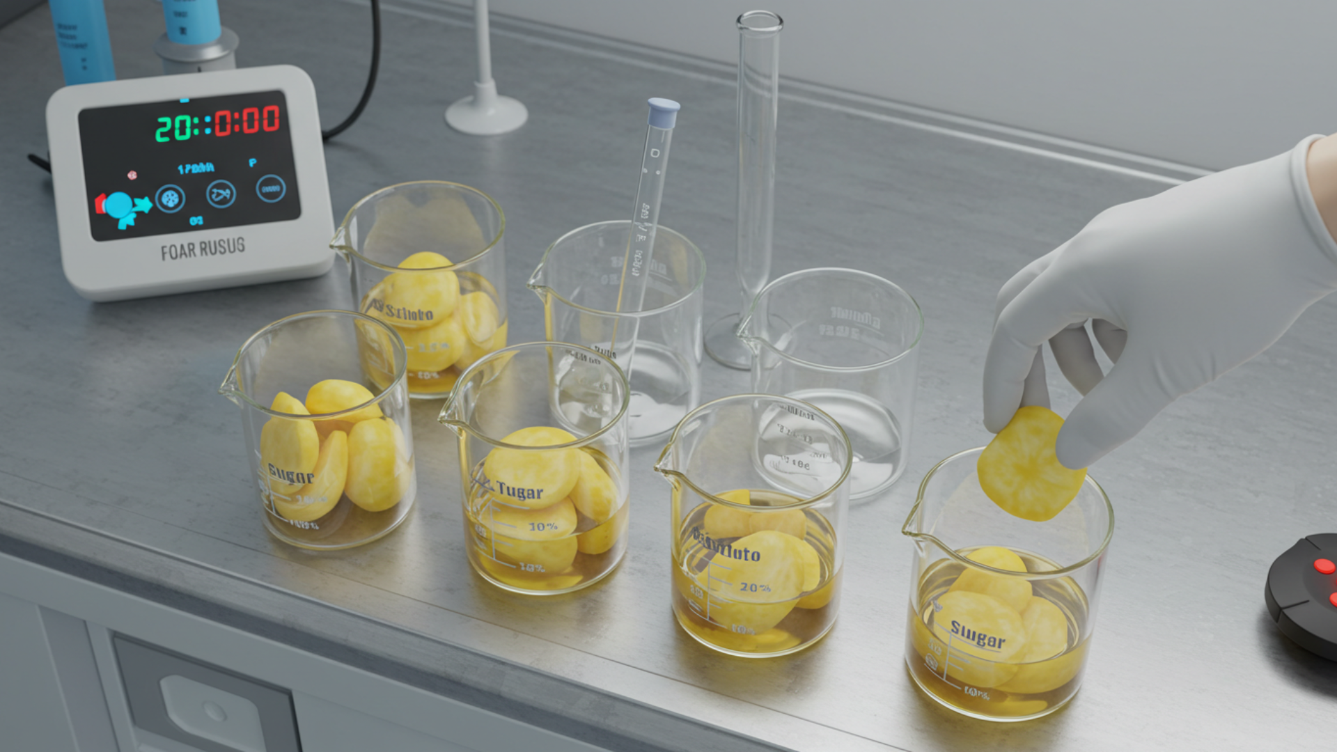
VR Plant Osmosis & Water Relations
We welcome your questions, do not hesitate to contact us
- Create virtual reality and augmented reality experiences at the best prices
- Equipping engineering laboratories with the best equipment
- Virtual tours with 360° technology and also feature that you can see them through just one link
Why partner with us?
- Immersive, interactive VR labs that boost learner engagement.
- Safe, repeatable simulations without fragile equipment.
- Fast iterations to match your curriculum and objectives.
Client & Visitor Reviews
Share your experience, feedback, or opinion about this page content.
Write a Review
Latest Reviews